
Must Read for Morgellons Patients and Caregivers
"I highly recommend this book for any patient suffering to educate themselves and
gain hope. It will also be an invaluable tool for loved ones of those who suffer,
so they can better grasp the reality of Morgellons Disease. Finally, medical
professionals can gain an understanding of what is involved in the diagnosis and
medical management of this complex disease. Patients are not delusional and
deserve care. Thank you Dr. Savely for bravely speaking up for those of us who
are discounted by mainstream medicine." -Molly Hasty, Amazon reviewer

Read Dr. Ginger Savely's book on Morgellons disease
(Paperback, 2016, Amazon.com)
WHAT IS MORGELLONS?
Morgellons disease is a system-wide illness specifically characterized by the presence of filaments of different colors underneath and protruding from the skin causing skin eruptions, pain and terrible itching. The disease has been associated with spirochetal bacteria, particularly the Borrelia spirochete that causes Lyme disease. Although, research is beginning to show the undeniable physiological basis to the disease, health care providers continue to insist that the victims are delusional and self-mutilating, compounding the suffering experienced by these unfortunate souls.
FOREWORD TO THE BOOK
There is a rapidly growing interest in Morgellons disease as evidenced by the fact that research papers on the topic have been among the most highly accessed of all scientific research on the Internet for the past few years. As director of the Charles E. Holman Morgellons Disease Foundation ( CEHMDF ), I hear from 5 to 10 new patients daily who have symptoms consistent with the disease and who have hit brick walls trying to access medical care.
In our early years, the CEHMDF would field calls from hostile doctors, disgruntled that we were validating the symptoms reported by Morgellons patients. Today we hear more often from doctors who are baffled and at a loss to know what to do for the patients presenting to their practices with the debilitating symptoms of this new and misunderstood disease. As more and more research is published in the medical literature, Morgellons disease is slowly gaining acceptance as an infectious disease despite an unusually fierce opposition that strives to keep it tucked away and dismissed as purely psychiatric in origin.
Dr. Ginger Savely was the first pioneering medical provider to take her Morgellons patients' symptoms seriously and work to legitimize the disease. She took on this very challenging patient population and stood by them in their darkest hours. Dr. Savely listened to her patients and looked at the blatant objective evidence. She was the first to speak out publicly about this serious health crisis and she has continued throughout the years to stand up for the reality of Morgellons disease through her numerous published articles, interviews, and presentations at medical conferences. Dr. Savely has been a lifeline for hundreds of suffering people who have lost their way and given up hope of ever receiving medical attention for their unusual and agonizing symptoms. She has worked to educate her peers and has been committed to helping her patients.
At a time when interest in Morgellons disease is at an all time high, this detailed first- hand account of the Morgellons journey is a much-needed resource. With clarity, personal style and years of experience, Dr. Savely has taken on a challenging and highly controversial subject. Using up-to-the-minute scientific findings, color photographs and sweeping examples she has illuminated the path to recognition of this debilitating and disfiguring disease, previously orphaned by the medical community.
We have come a long way against great odds on our journey to legitimize Morgellons disease. Dr. Savely paved the earliest path on the rockiest road to make this progress possible. I have no doubt that Morgellons disease will eventually be fully recognized worldwide by health agencies and the medical community at large because the scientific evidence has reached an undeniable point. I firmly believe that in time the truth will prevail.
This book provides long-awaited answers to the vital questions being posed by thousands of patients and clinicians about this unthinkable disease. In reading Dr. Savely’s poignant narrative, you will learn to separate fact from myth and rumor from reality. This book will be a valuable resource for Morgellons patients and their families and friends and will serve as a guide for clinicians who are open-minded and committed to the health of their patients.
Cindy Casey-Holman, RN
Director The Charles E. Holman Morgellons Disease Foundation
www.MorgellonsDisease.org
“Never doubt that a small group of thoughtful,
committed citizens can change the world;
indeed it's the only thing that ever has.”
— Margaret Mead
COLOR IMAGES
OF MORGELLONS DISEASE PATIENTS
TO ACCOMPANY THE BOOK
Click to enlarge
Image 1: Morgellons disease (MD) patients often present to the office looking like this.
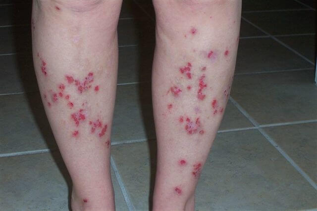
Image 2: Lesion with black filaments.

Image 3: Lesions on scalp with hair loss.
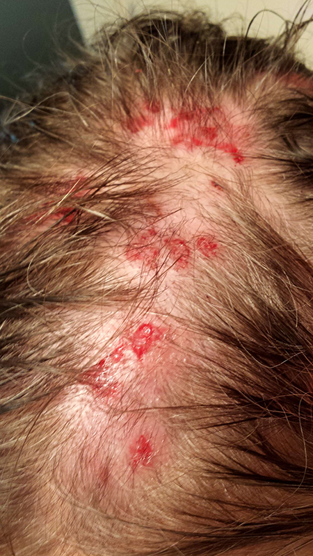
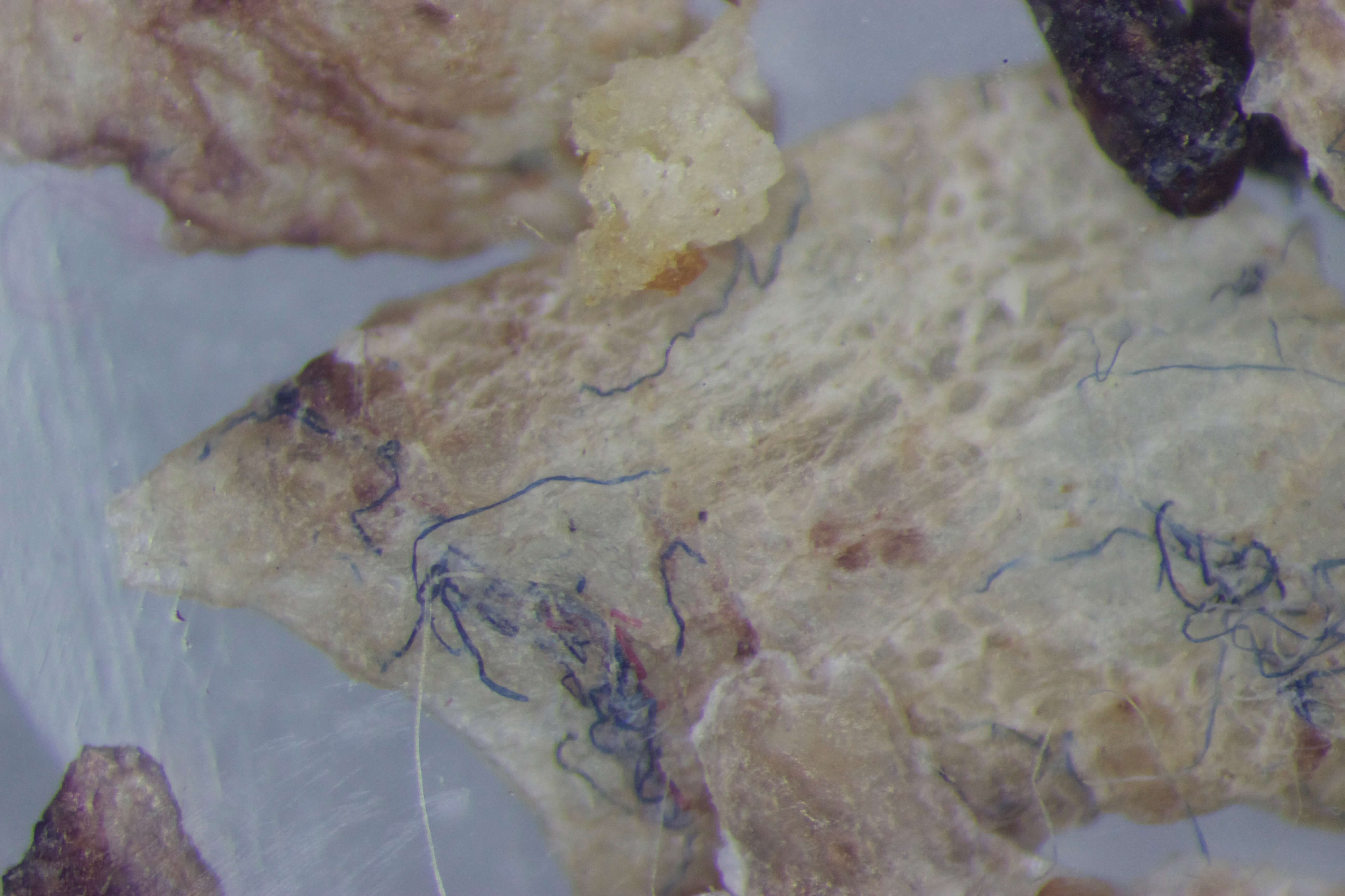
Image 4: Blue and white filaments seen intertwined in a scab.
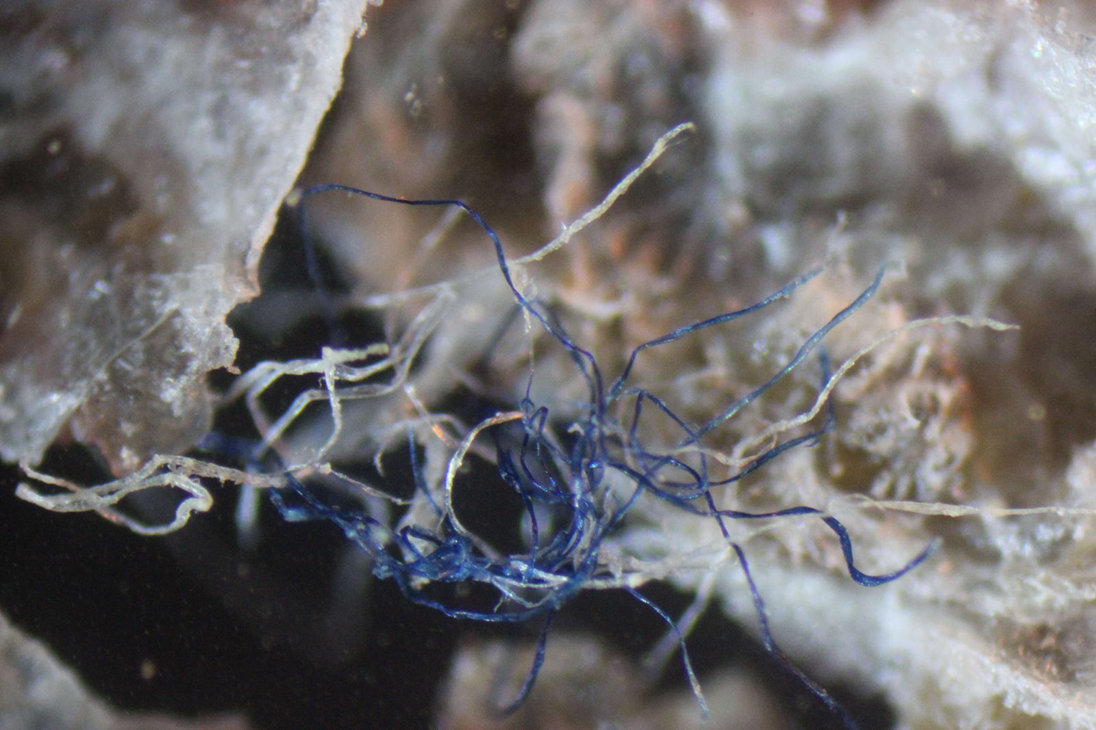
Image 5: Black filaments in a scab.
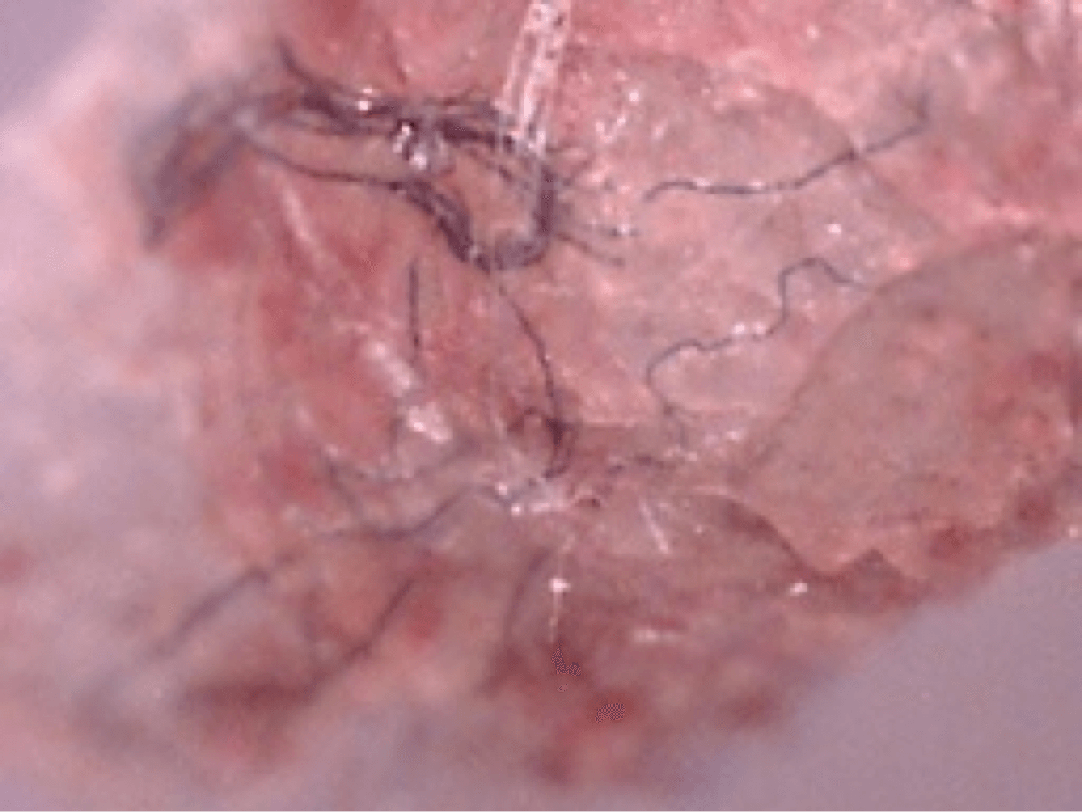
Image 6: Blue filaments in a scab.
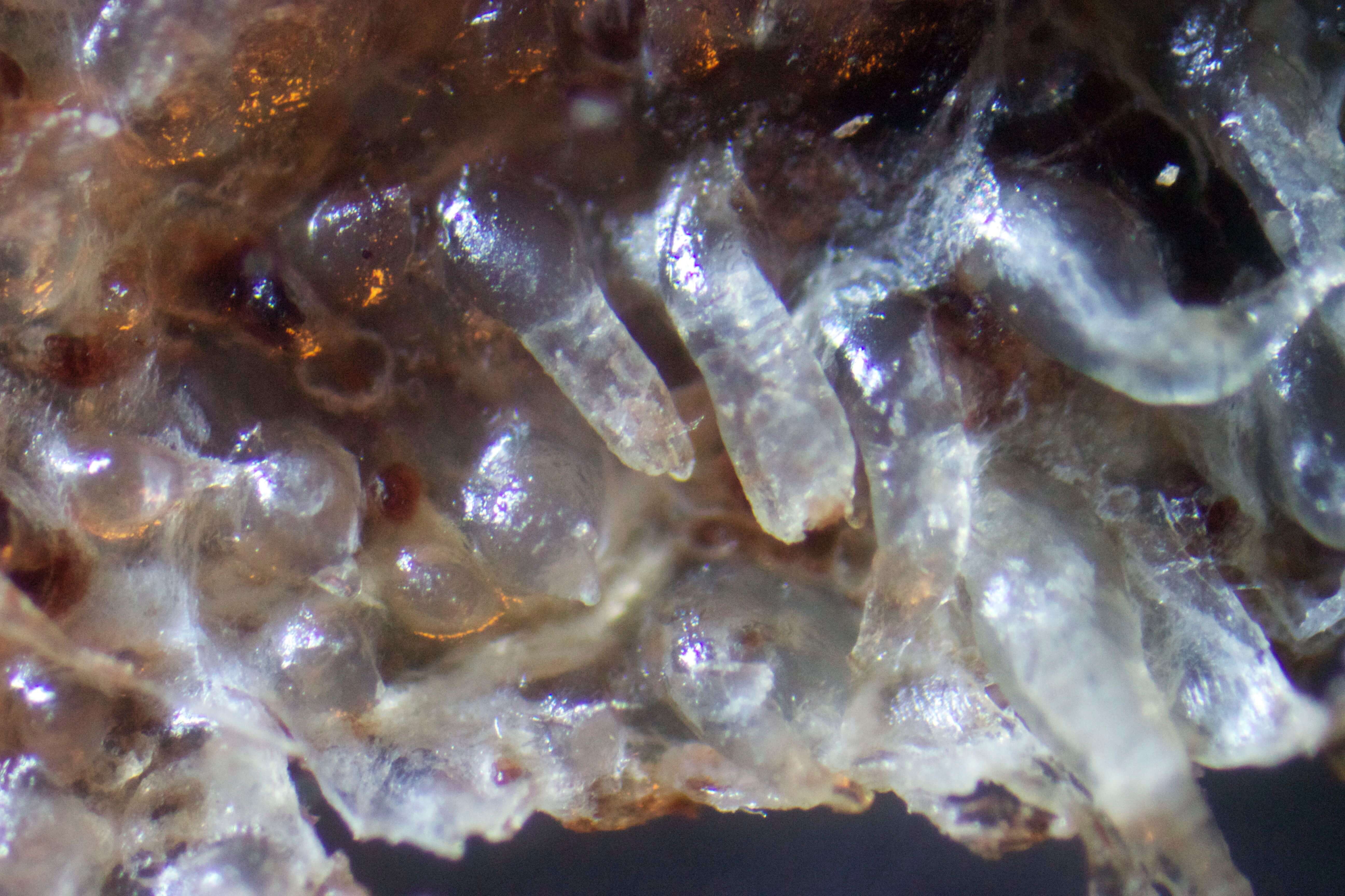
Images 7 and 8: Follicular casts seen at 100x magnification on the underside of removed calluses from MD patients.
Images 9 and 10: Although these scabs showing keratin “plugs” were removed from one of my patients, they look very much like those seen in cows with Bovine Digital Dermatitis.
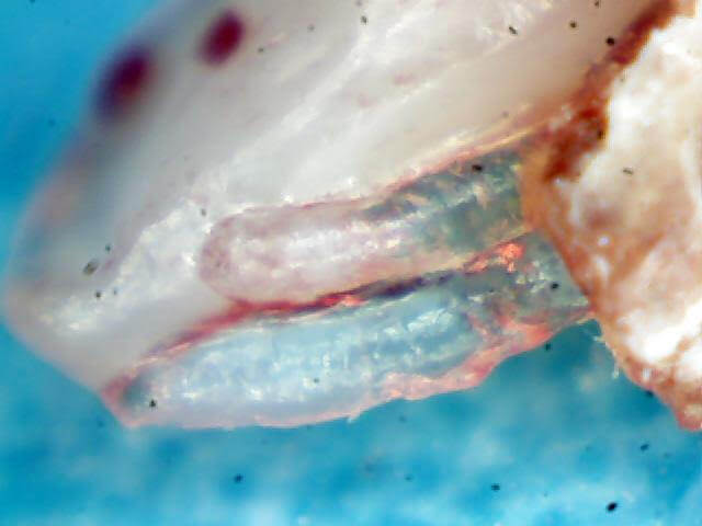
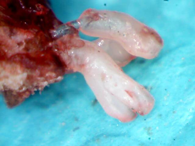
Image 11: A scab with keratin projections underneath as well as a filament that was growing down into the dermis.
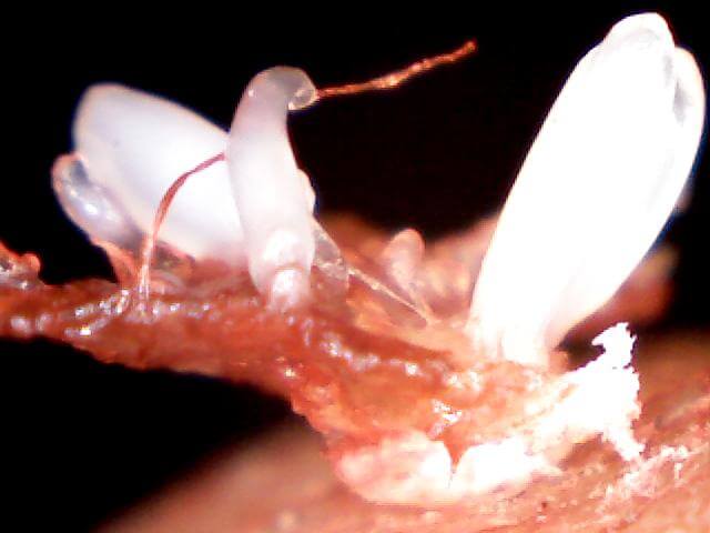
Image 12: Example of a deep, circular lesion that some MD patients have.

Image 13: Same patient as above with worsening of deep lesions.
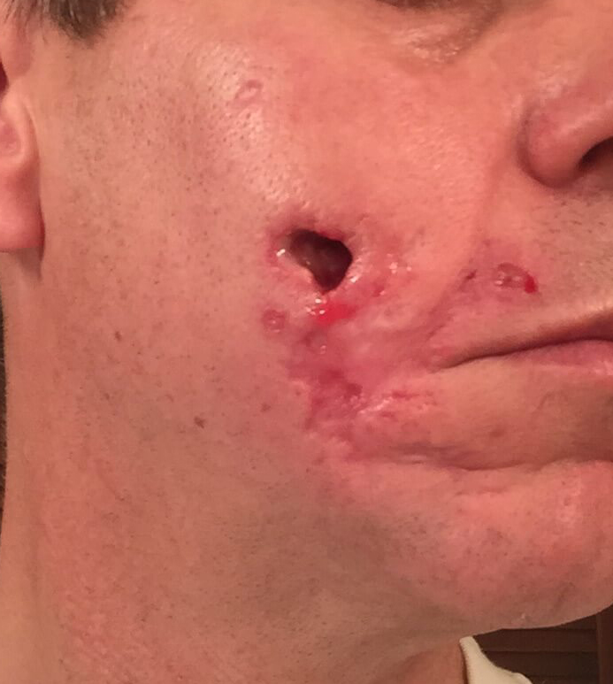
Image 14: Same patient as above.
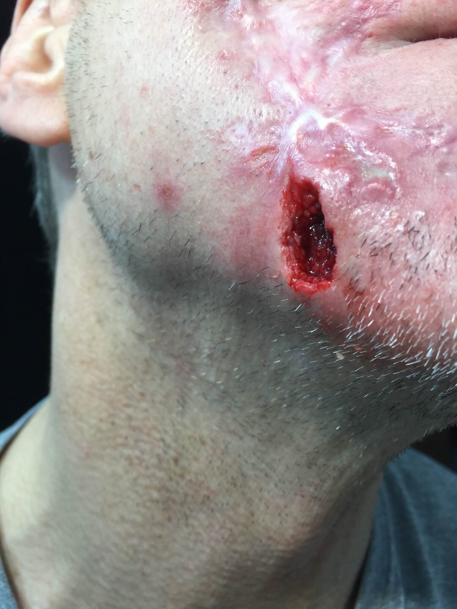
Image 15: Same patient. Deep lesions continue forming on the right side of his face.
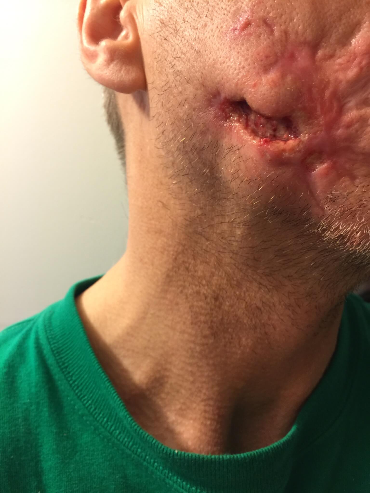
Image 16: Same patient after his numerous facial lesions had temporarily healed. Later his face opened up again in several new places.
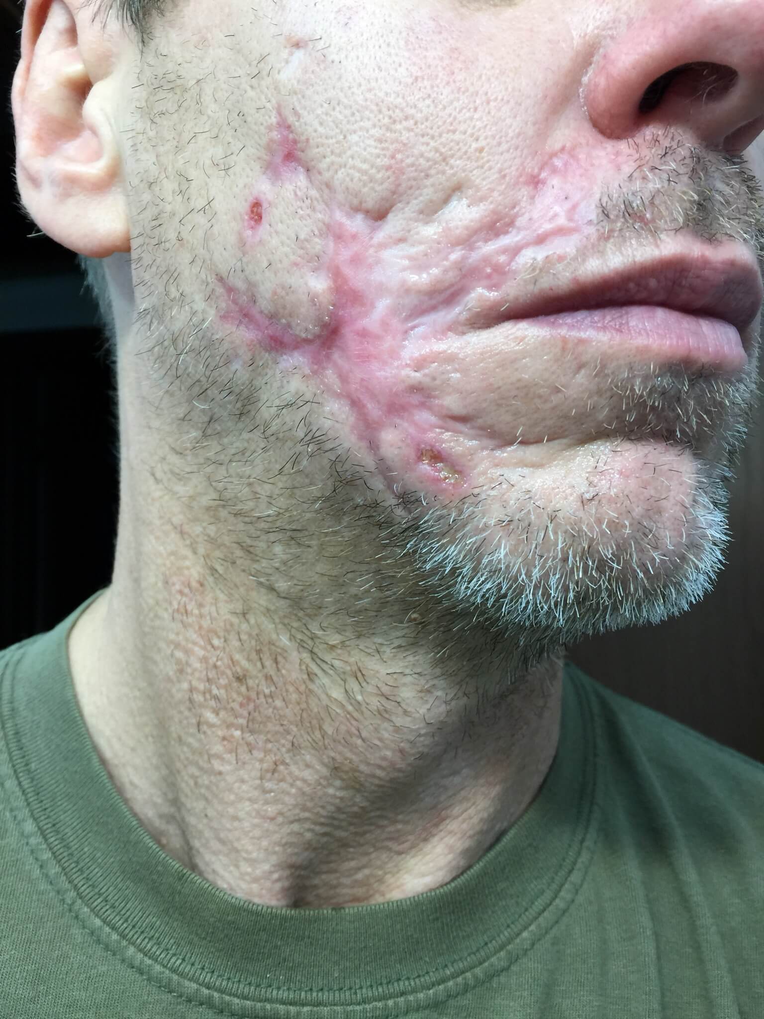
Image 17: Testicle of one of my patients as another example of the deep
lesions some MD patients have.
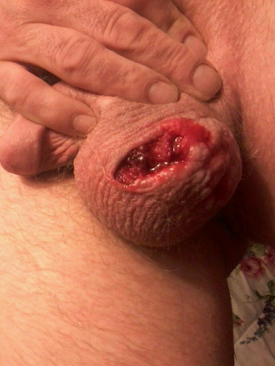
Image 18: Black and red filaments are seen in the web between a patient’s thumb and forefinger. This photograph is on the book’s front cover.
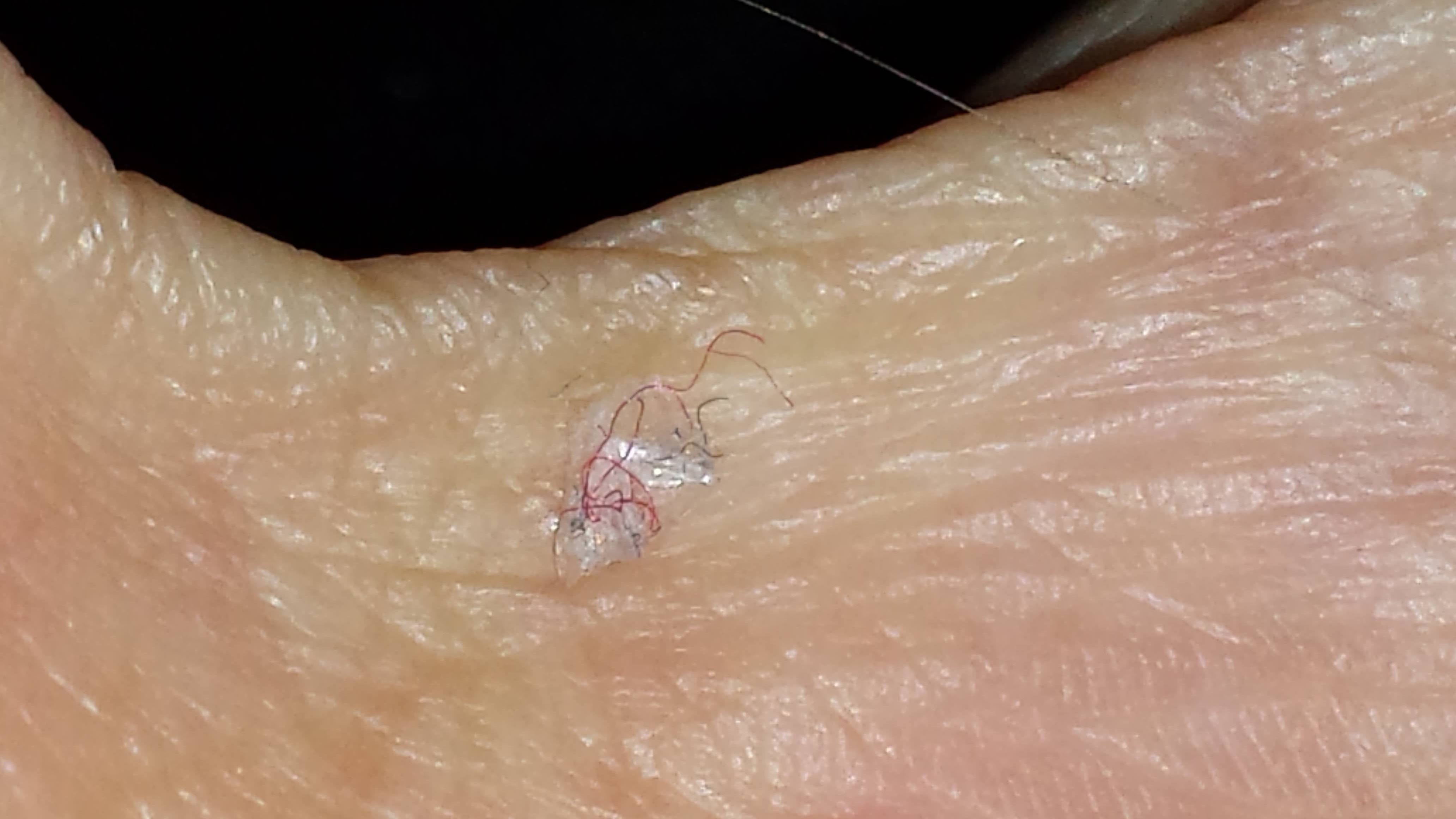
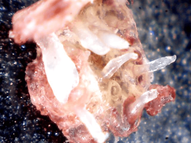
Image 19: Clear to white filaments are often seen growing from the underside of nails of MD patients.

Image 20: Black and white filaments as seen under the epidermis at 200x.
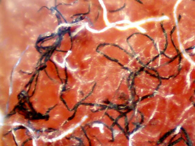
Image 21: A flat ribbon-like filament, this one from a vagina.
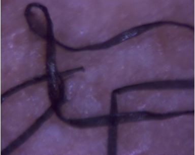
Image 22: An example of a filament that looks like a feather.
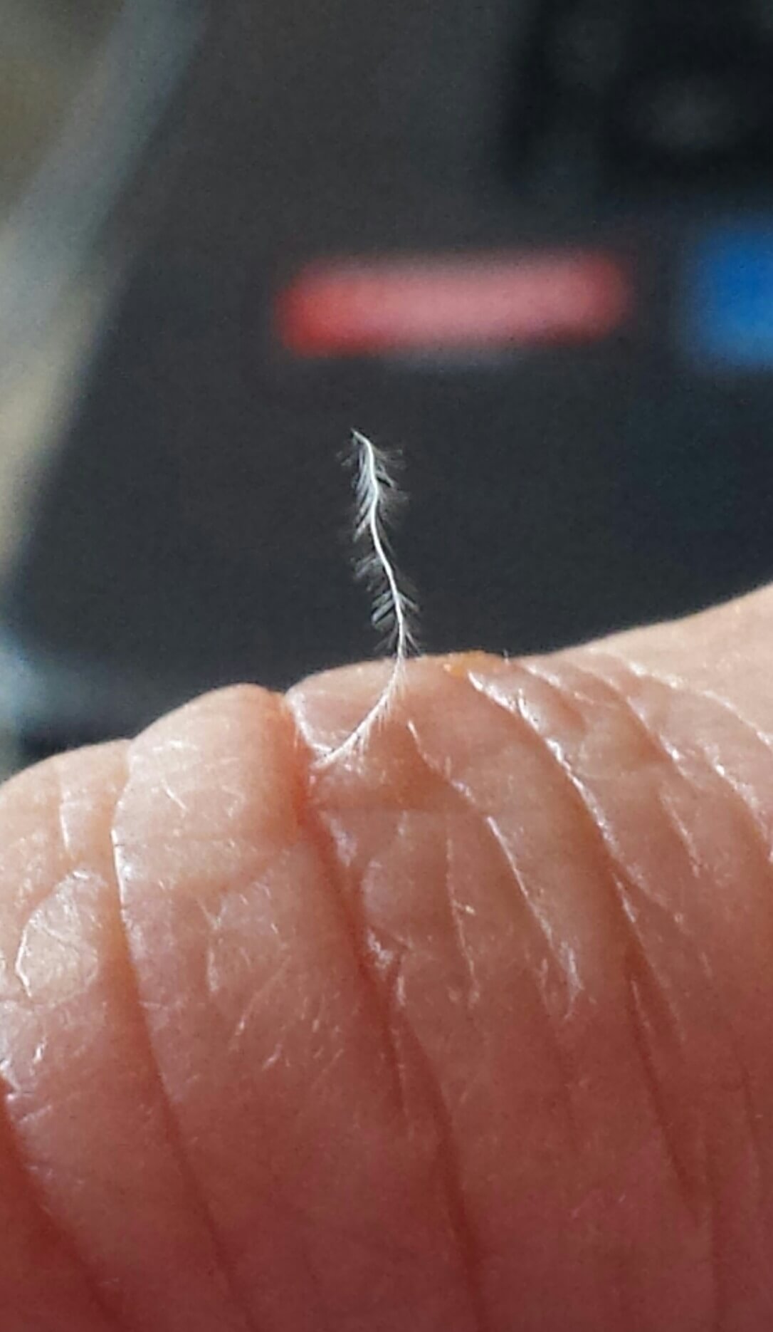
Image 23: Another feather-like filament.

Image 24: Keratin "shard" that was deeply embedded in an MD patient's lesion.
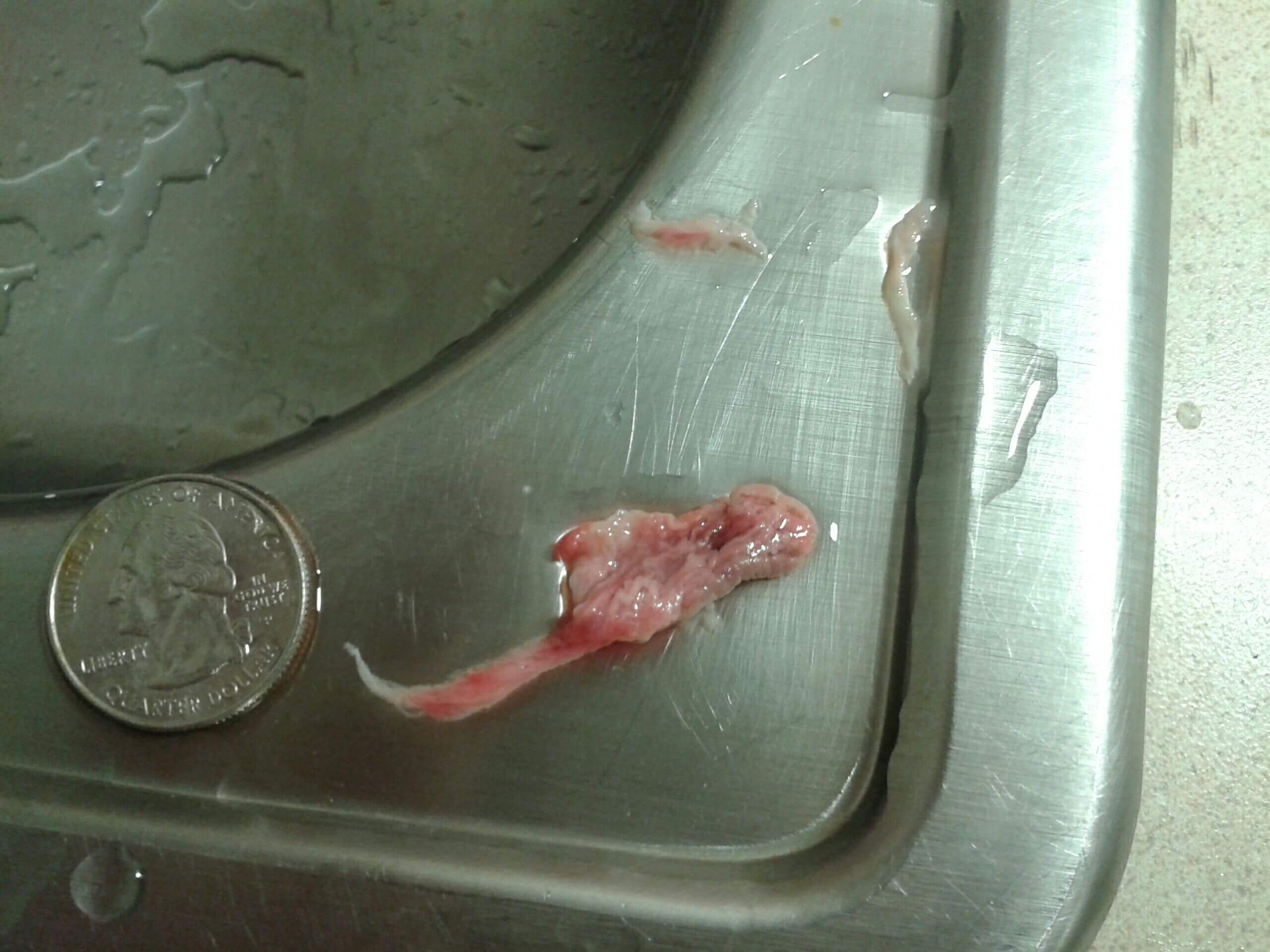
Image 25: Another keratin "shard".
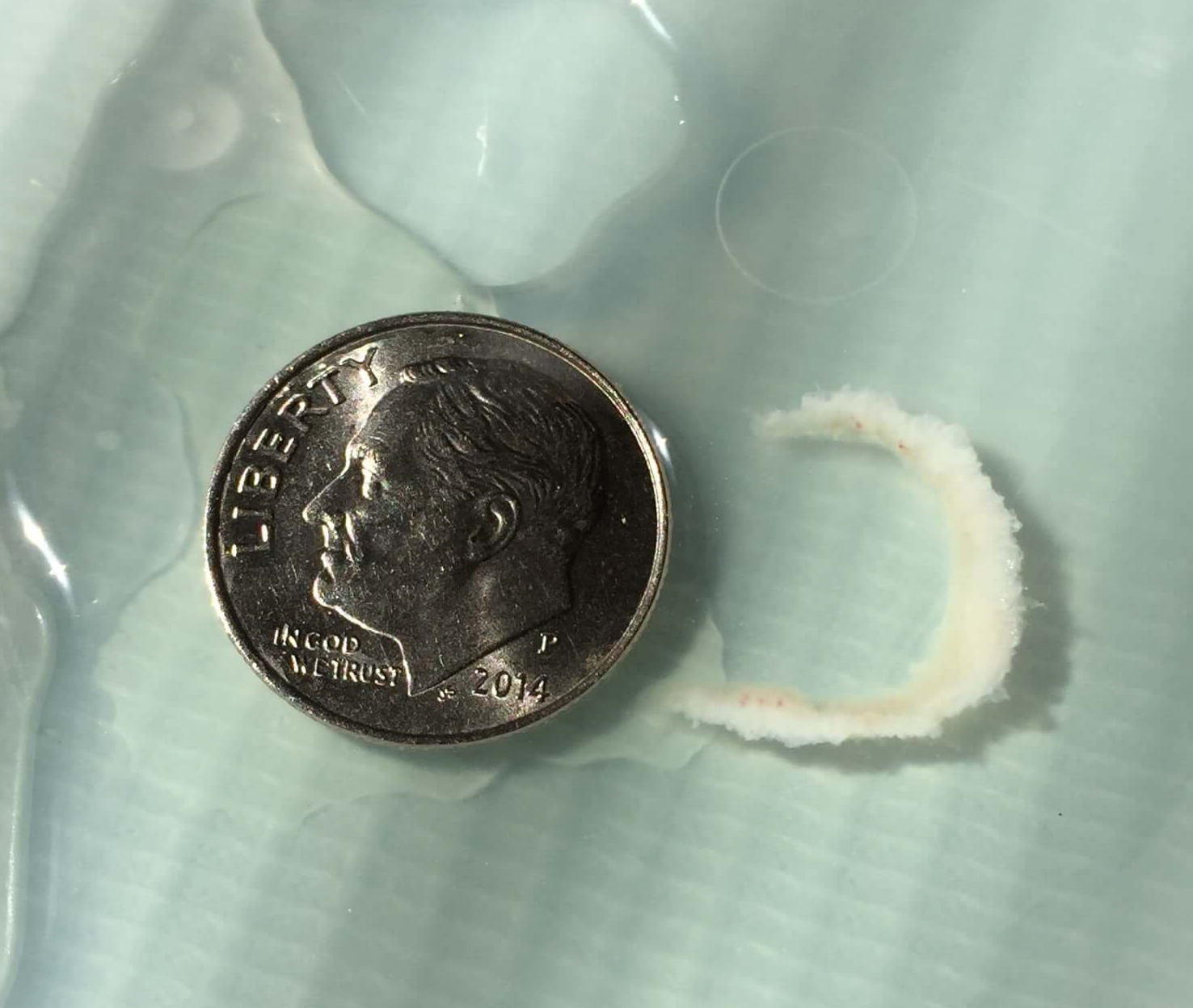
Image 26: Another keratin “shard”.
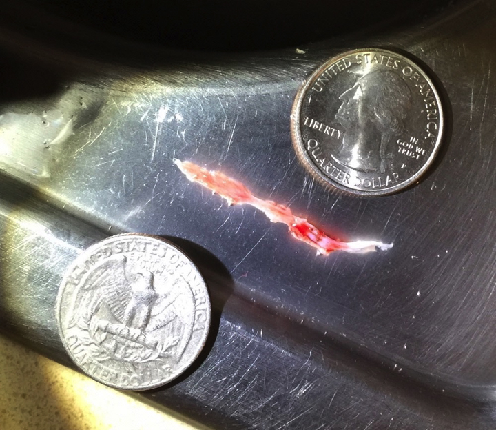
Image 27: A black speck amidst fibers in a skin sample from an MD patient. 70x magnification. Courtesy of M. Middelveen.
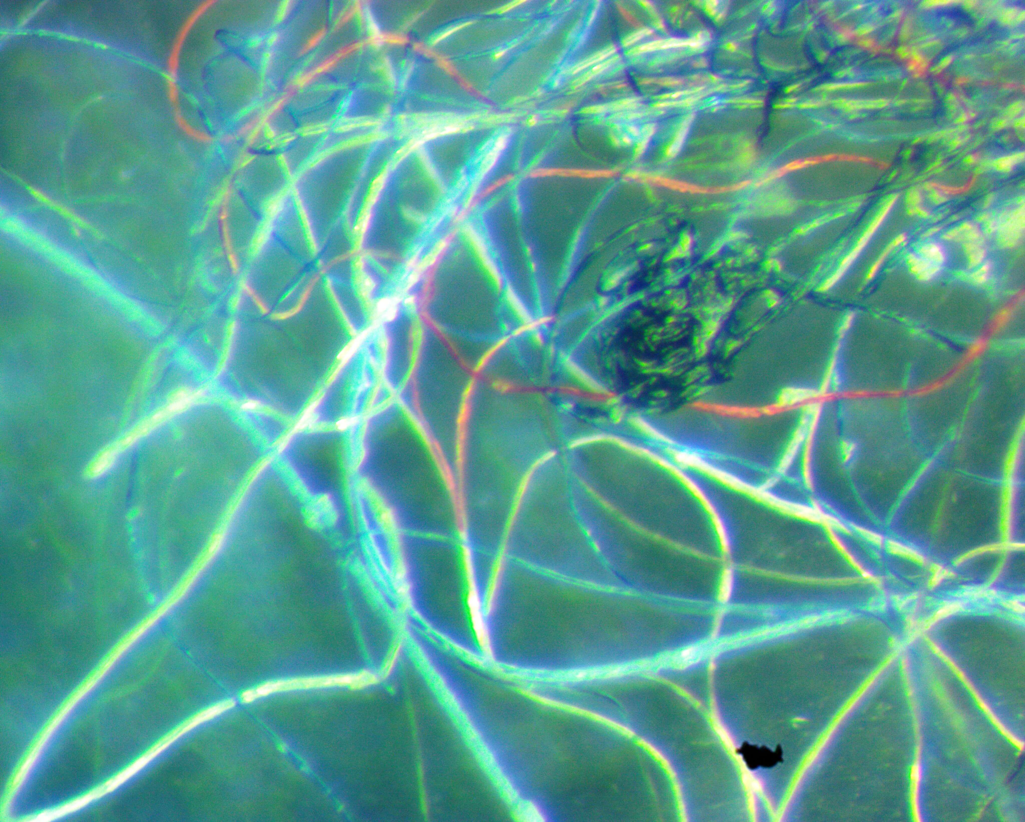
Image 28: A mass of black fibers (patients refer to these as "fuzz balls")
at 60x magnification.
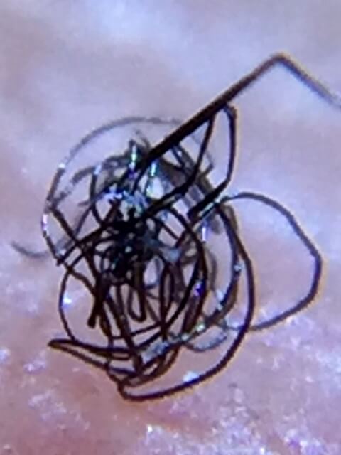
Image 29: Red, blue and white filaments in the gelatinous material that MD
patients complain of.
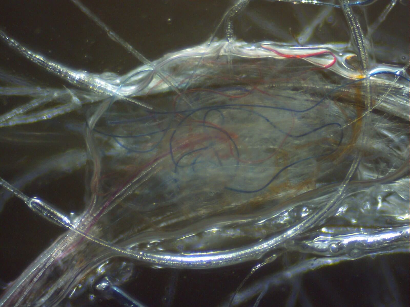
Image 30: Single gelatinous "plug" seen on the underside of a small scab
removed by a nurse with the CEHMDF.
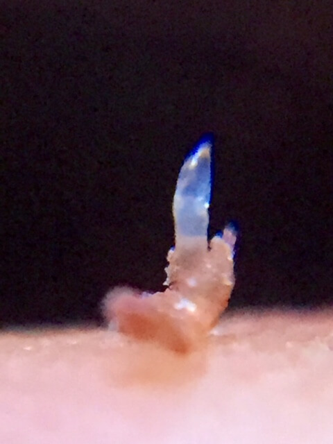
Image 31: Thick, black liquid that comes out of the pores of about 30% of MD patients.
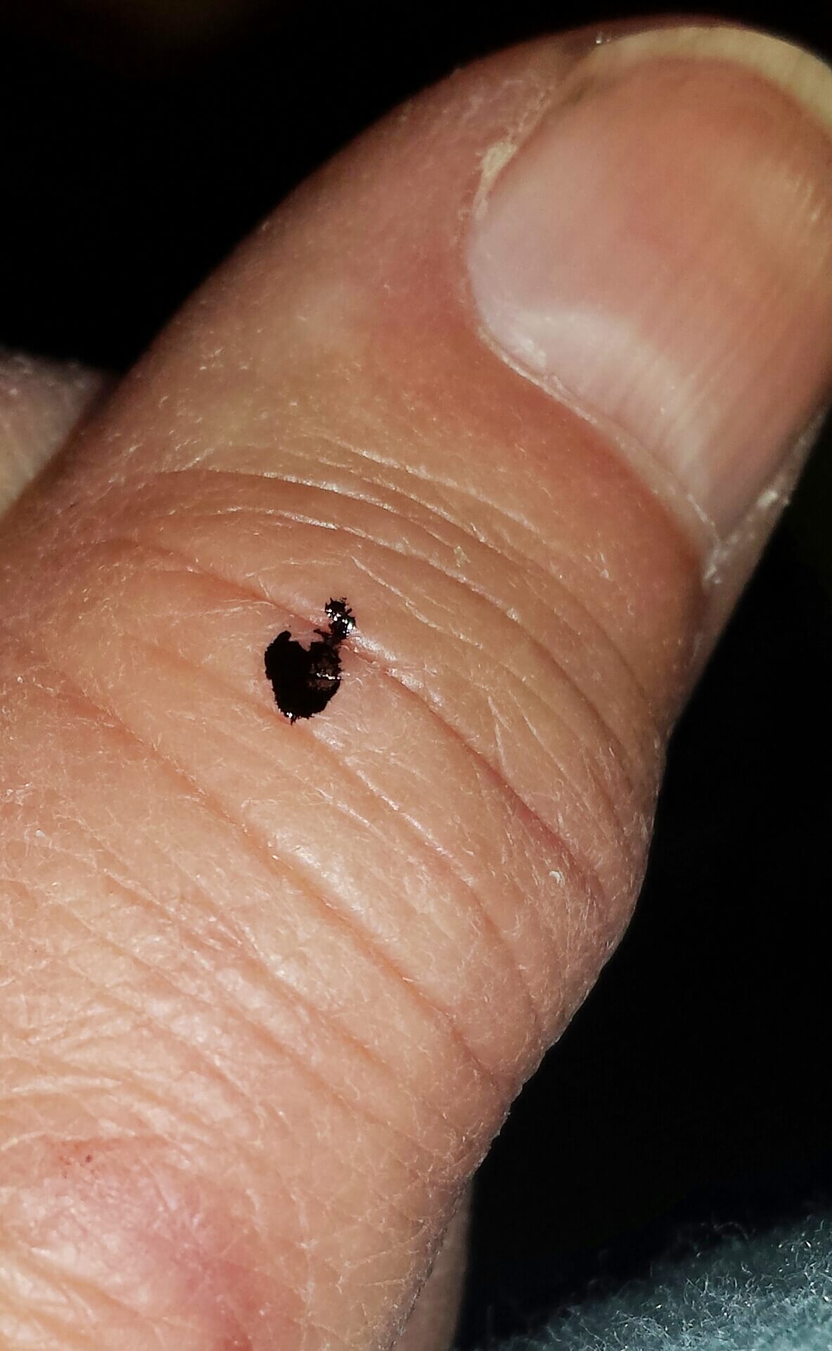
Image 32: Crystal-looking exudates that many MD patients report.
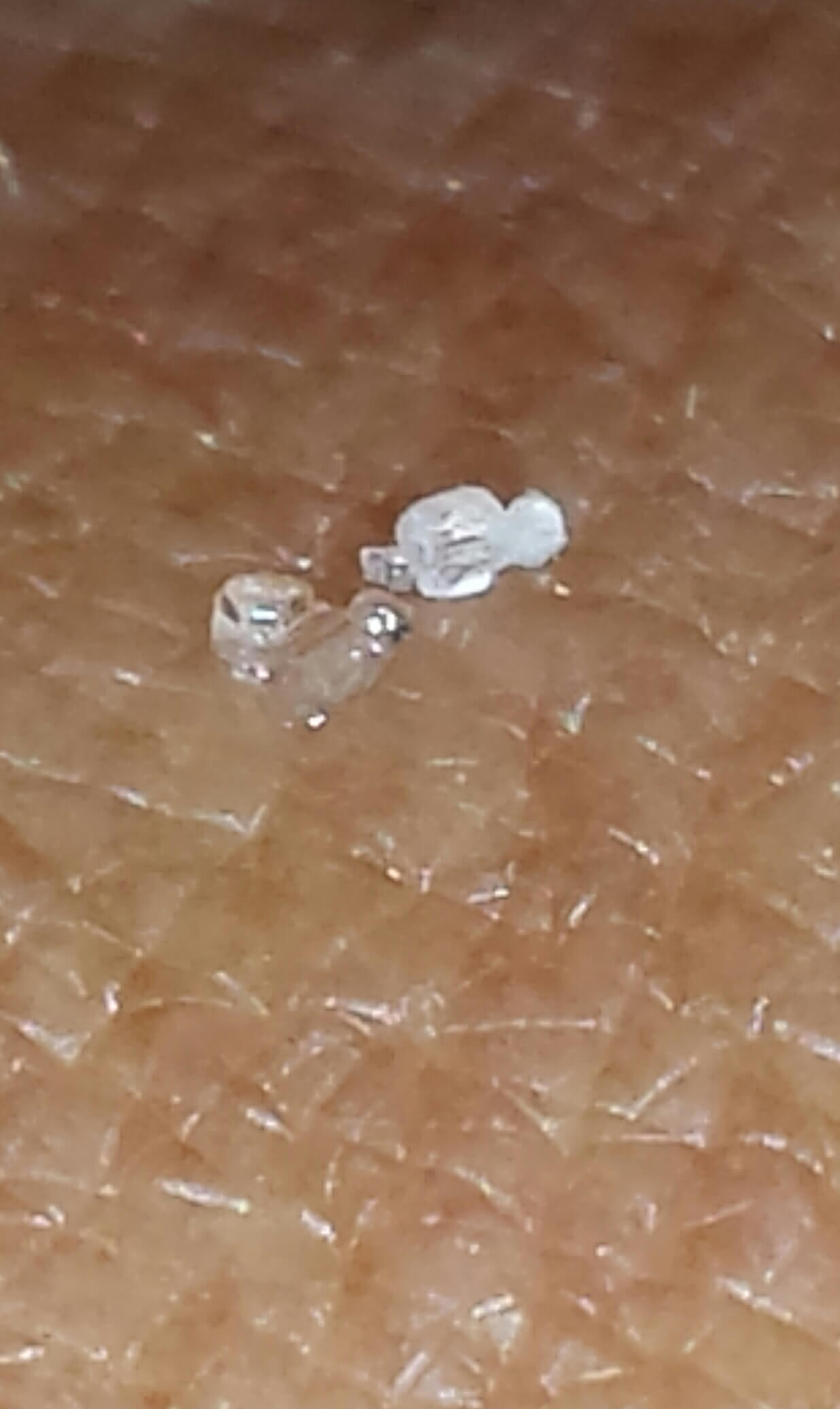
Image 33: Filaments tightly wound around a human hair at 200x.

Image 34: Blackening of teeth close to the gum line. Photo courtesy of Katie Yussuf.
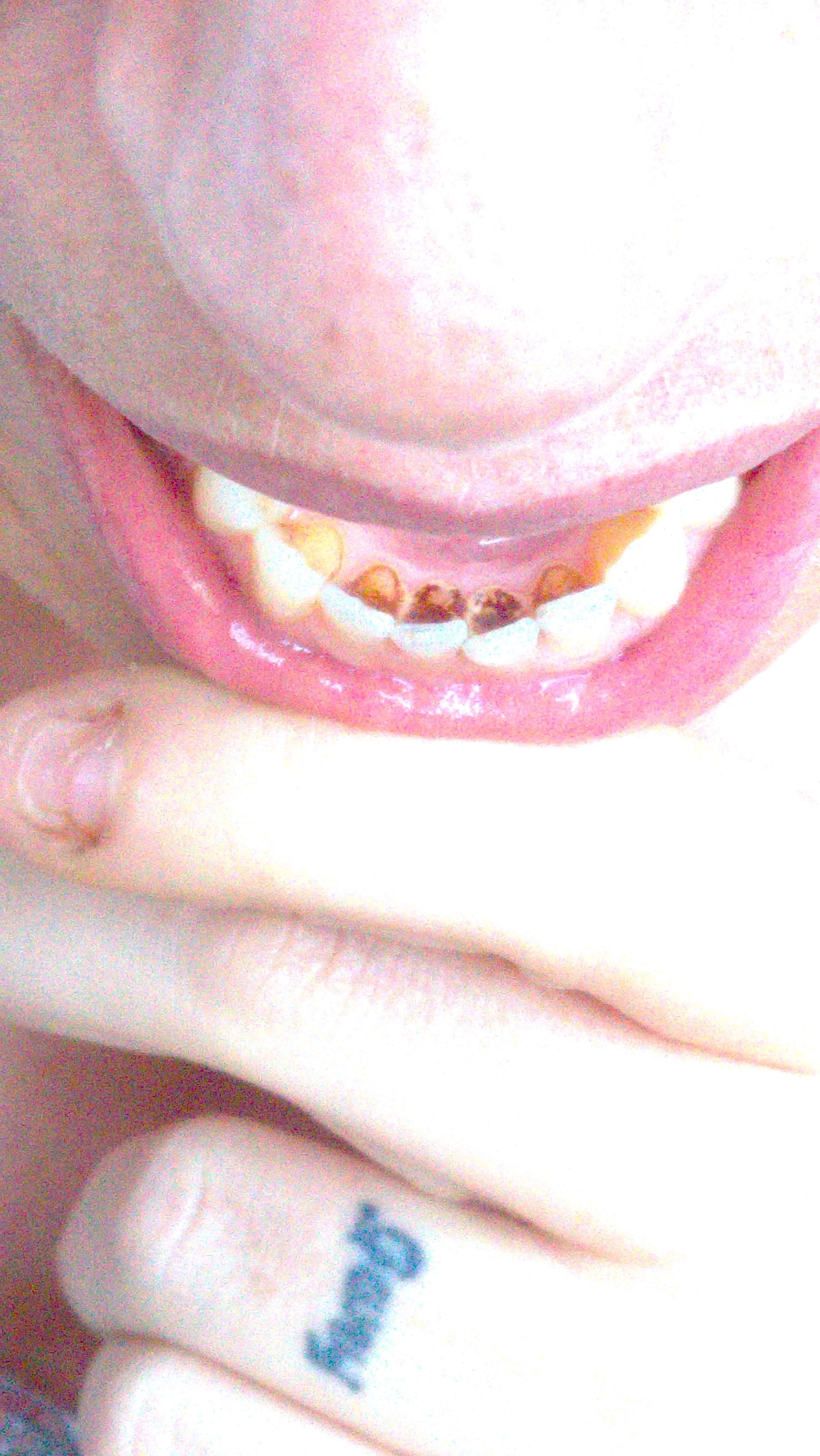
Image 35: Example of the spontaneous "scratches" that appear on the skin of many MD patients.
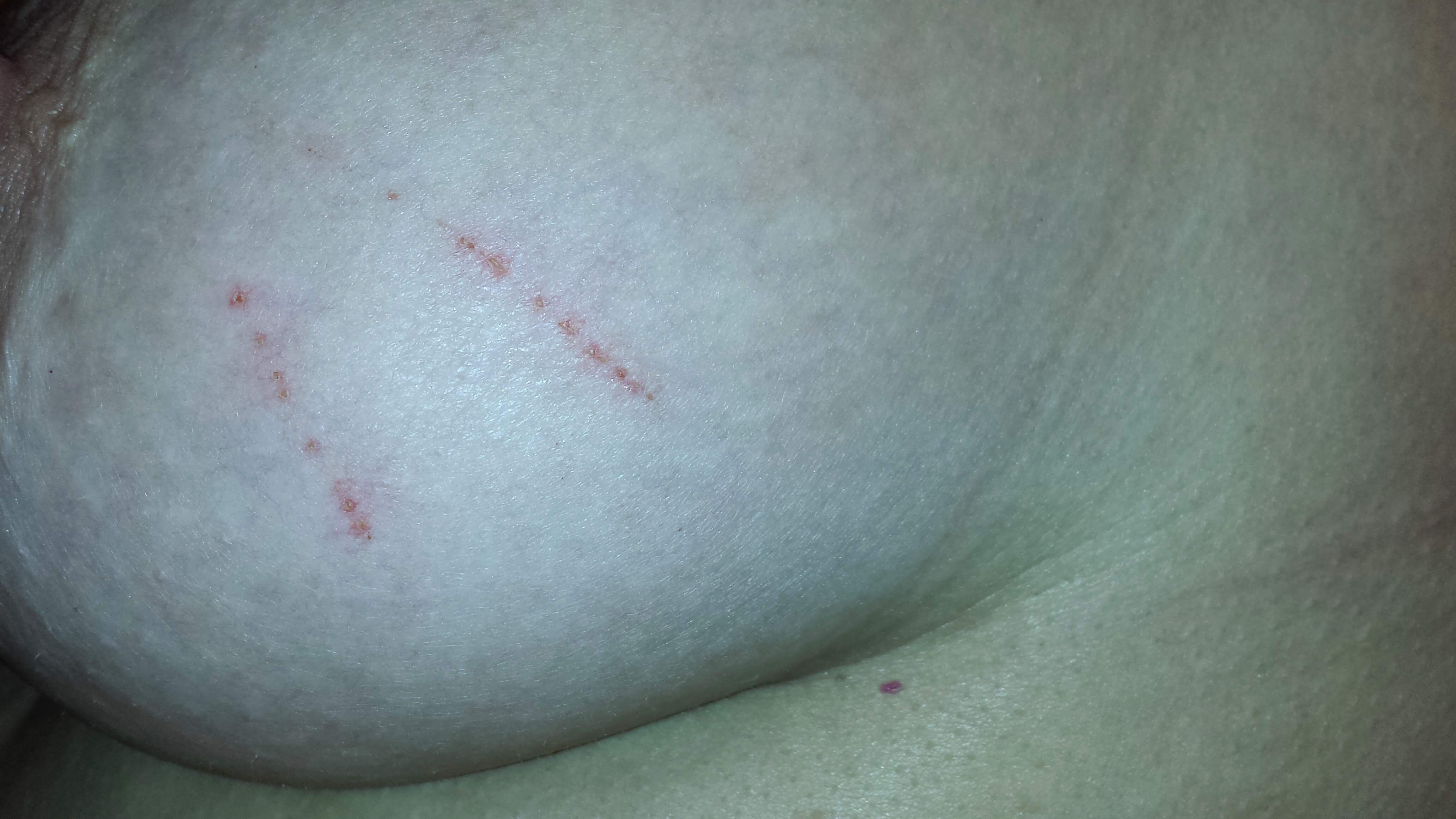
Image 36: Streaks on an MD patient’s skin consistent with Bartonella striae.

Image 37: Linear, raised "tunnels" visible on the skin of many MD patients.
These are on a patient's knee.
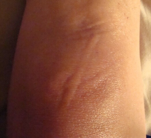
Image 38: These "tunnels" are on a breast.
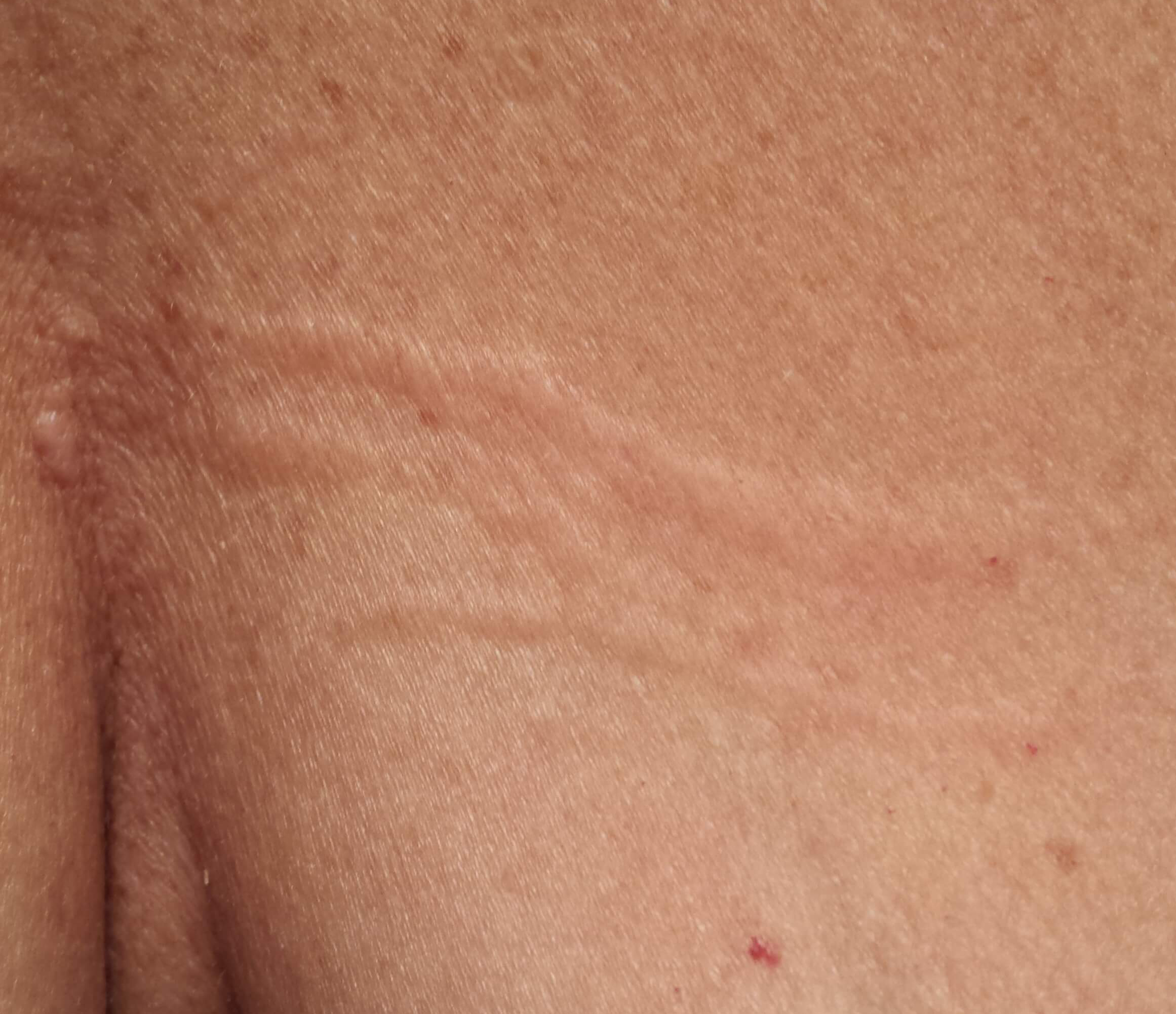
Image 39: Filaments tightly wind around follicular sheaths in the hair
and are often mistaken by patients as "cocoons".
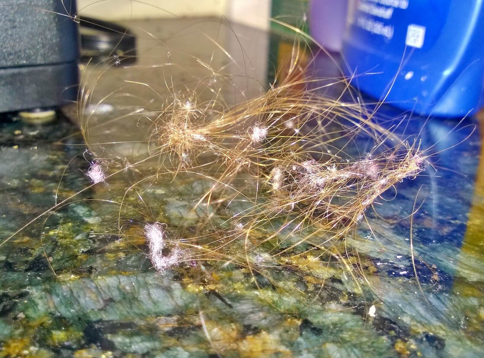
Image 40: Mound on crown of shaved head.
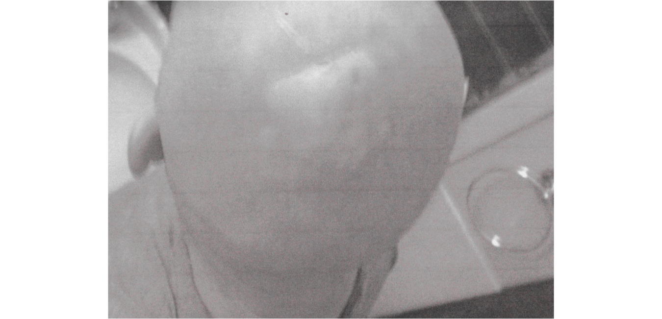
Image 41: Mound with lesions, showing loss of hair in that region.
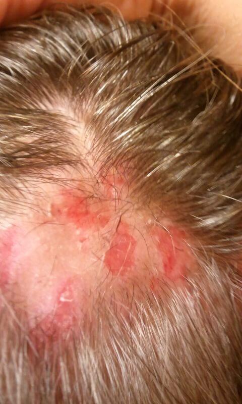
Image 42: MD patient skin sample stained with Gomori Trichrome stain which
turns keratin red and collagen a bluish-green.
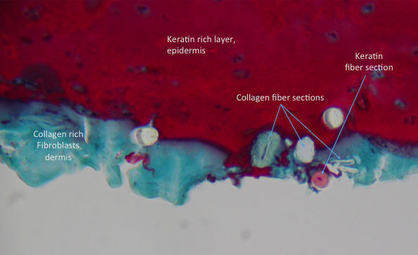
Image 43: 500x magnification. Spirochetal structures seen in MD patients'
skin lesions using fluorescent immunohistochemical analysis with
Borrelia-specific monoclonal and polyclonal antibodies. (Photo courtesy
of Eva Sapi, Ph.D.)
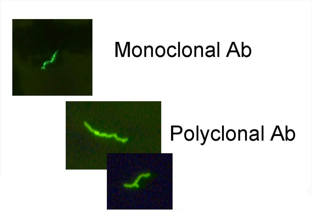
Image 44: Photomicrograph of a spirochete (in this case, Treponema pallidum), by Bill Schwartz, CDC, Courtesy: Public Health Image Library. Public domain.

Image 45: Dog with MD lesions. Photo courtesy of Edward C. Rasmussen.

Image 46: Dog whisker with gelatinous material at the root, similar to what is seen in humans with MD. Photo courtesy of Lee M. Laskowsky.
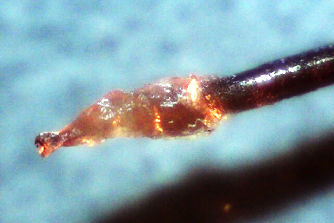
Image 47: Leg of patient before treatment.
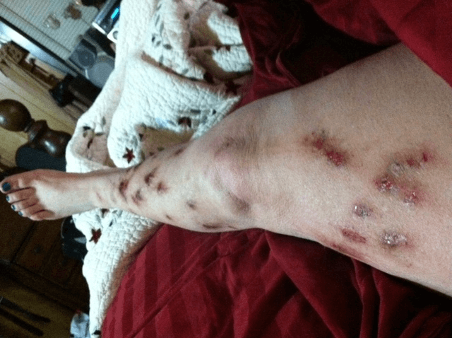
Image 48: Legs of same patient after treatment.